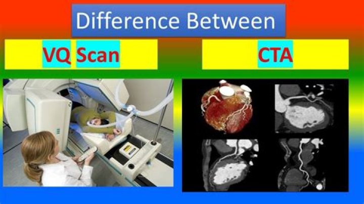
A ventilation–perfusion (VQ) scan is a nuclear medicine scan that uses radioactive material (radiopharmaceutical) to examine airflow (ventilation) and blood flow (perfusion) in the lungs. The aim of the scan is to look for evidence of any blood clot in the lungs, called pulmonary embolism (PE)..
Regarding this, how long are you radioactive after a VQ scan?
You need to stay very still during the scans to avoid blurring the pictures. Afterward, the radioactive gas or mist will clear from your lungs as you breathe. The ventilation scan takes about 15 to 30 minutes.
is dye used in a VQ scan? A VQ scan (ventilation-perfusion lung scan) is a nuclear medicine imaging study. VQ scans can be used to help diagnose pulmonary embolism in patients who cannot receive iodinated contrast (X-ray dye), such as that used in computed tomographic angiography (CTA).
Beside above, when would you use a VQ scan?
A VQ scan is used most frequently to screen for a pulmonary embolus, which is also known as a blood clot in the lungs. Symptoms of pulmonary embolus may include: rapid heart rate. trouble breathing.
How accurate is a VQ scan?
Several papers have reported statistically significant greater accuracy for PE detection for CT with sensitivities and specificities for CT of 83% to 94% and 94% to 96%, respectively vs. 65% and 94% for V/Q scintigraphy.
Related Question Answers
Is a VQ scan dangerous?
What are the risks of a VQ Scan? There are minimal risks involved in the VQ scan. Allergic reactions to the radiopharmaceuticals are rare and will be treated as needed. The test involves exposure to ionising radiation (see Radiation Risk of Medical Imaging in Adults and Children).How much radiation is in a VQ scan?
Breast radiation estimates made using 4-slice CT vary from 20 to 60 mSv (4–6), whereas that from V/Q is approximately 0.28–0.9 mSv (7). A recent report by Einstein et al. (8) estimated that 64-slice chest CTA delivers a dose of 50–80 mSv to the breast.Can a VQ scan cause cancer?
The risk to your baby of developing childhood cancer after a VQ scan or a CT scan is slightly higher with a VQ scan. This risk is an extra 1 case of cancer for every 34,000 VQ scans performed.Can a VQ scan show pneumonia?
A V/Q lung scan may be performed in the case of serious lung disorders such as chronic obstructive pulmonary disease (COPD) or pneumonia as well as a lung performance quantification tool pre- and post-lung lobectomy surgery.What causes V Q mismatch in asthma?
A V/Q mismatch happens when part of your lung receives oxygen without blood flow or blood flow without oxygen. This happens if you have an obstructed airway, such as when you're choking, or if you have an obstructed blood vessel, such as a blood clot in your lung.Will a chest xray show a pulmonary embolism?
Chest X-ray This noninvasive test shows images of your heart and lungs on film. Although X-rays can't diagnose pulmonary embolism and may even appear normal when pulmonary embolism exists, they can rule out conditions that mimic the disease.Can you have a pulmonary embolism with a normal D dimer?
Background: d-dimer testing is commonly used to help exclude venous thromboembolism (VTE), such as pulmonary embolism and deep venous thrombosis (DVT). Patients with low clinical probability and normal d-dimer levels of 0.5 mcg per L (0.5 mg per L) or less had pulmonary embolism ruled out.What can a pulmonary function test diagnose?
Pulmonary function tests, or PFTs, measure how well your lungs work. They include tests that measure lung size and air flow, such as spirometry and lung volume tests. Other tests measure how well gases such as oxygen get in and out of your blood. These tests include pulse oximetry and arterial blood gas tests.What happens during a VQ scan?
A lung VQ scan is an imaging test that uses a ventilation (V) scan to measure air flow in your lungs and a perfusion (Q) scan to see where blood flows in your lungs. It uses special x ray scanners outside of your body to create pictures of air and blood flow patterns in your lungs.How long does a lung scan take?
The camera does not produce any radiation, it simply detects and records the distribution of the radioactive material in your lungs. This part of the test will take about 15 to 20 minutes. For the second part of your test, you will be asked to lie flat on your back on the examination table.How is a CTPA performed?
CT pulmonary angiogram (CTPA) is a medical diagnostic test that employs computed tomography (CT) angiography to obtain an image of the pulmonary arteries. Its main use is to diagnose pulmonary embolism (PE). Images are acquired with the maximum intensity of radio-opaque contrast in the pulmonary arteries.How is a lung scan done?
A lung scan is a type of nuclear scanning test. It uses a special camera to take pictures of the lungs after a radioactive tracer is put into the body. It is most often used to find a pulmonary embolism. This is a blood clot that prevents normal blood flow in the lung.What is a D dimer test?
A D-dimer test is a blood test that can be used to help rule out the presence of a serious blood clot. But you can get high levels of D-dimer in your blood if you have a major clot like with deep vein thrombosis (DVT).What is perfusion in the lungs?
Gas exchange occurs in the lungs between alveolar air and blood of the pulmonary capillaries. Ventilation (V) refers to the flow of air into and out of the alveoli, while perfusion (Q) refers to the flow of blood to alveolar capillaries.How is ventilation perfusion scan done?
During the perfusion scan, a health care provider injects radioactive albumin into your vein. You are placed on a movable table that is under the arm of a scanner. During the ventilation scan, you breathe in radioactive gas through a mask while you are sitting or lying on a table under the scanner arm.What is a pulmonary angiogram used for?
Pulmonary angiography is a test to see how blood flows through the lung. Angiography is an imaging test that uses x-rays and a special dye to see inside the arteries. Arteries are blood vessels that carry blood away from the heart.How do you prepare for a chest CT scan?
EAT/DRINK : If your doctor ordered a CT scan without contrast, you can eat, drink and take your prescribed medications prior to your exam. If your doctor ordered a CT scan with contrast, do not eat anything three hours prior to your CT scan. You are encouraged to drink clear liquids.What is a VQ scan and pregnancy?
PREGNANCY. Patient Information. What is a lung VQ scan? A lung VQ scan looks at the air supply and blood supply to the lungs. It determines the likelihood of having a pulmonary embolism, a blood clot in the lung.What does a nuclear medicine test show?
What is General Nuclear Medicine? Nuclear medicine imaging uses small amounts of radioactive material to diagnose, evaluate or treat a variety of diseases. These include many types of cancers, heart disease, gastrointestinal, endocrine or neurological disorders and other abnormalities.